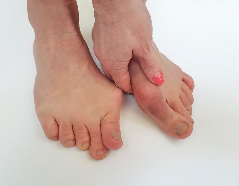
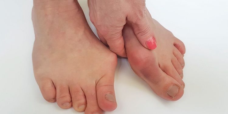
Hallux valgus symptoms include pain in the ball of your foot, a protruding bump on the inside of your big toe (hallux), and a change in how your big toe rolls when you walk. The bunion is formed when the bone at the front of your foot – the first metatarsal – is out of alignment with the other bones in the toe.
Symptoms can be mild or severe depending on the degree of the deformity and how it affects your lifestyle. Those with advanced bunion syndrome have significant pain when walking and may find it difficult to walk. They are also more likely to develop painful bursitis or arthritis in the big toe joint, called hallux rigidus.
In the early stages of hallux valgus, pain can be controlled by wearing special shoes that accommodate the bunion. These shoes provide more room for the bunion and reduce friction to minimize swelling and inflammation.
If a bunion is not corrected, the foot will continue to deform over time. This can cause a number of other problems, including arthritis and swollen joints.
When the metatarsophalangeal joint is misaligned, pressure is placed on nearby bones and tendons. This can cause swelling and redness, and can lead to pain.
Another common symptom is pain in the smaller toes, known as metatarsalgia. This is caused by the shift of weight from the big toe to the small toes when walking. The bursa in the ball of the toe can also become irritated and inflamed, which can result in pain and redness.

X-rays are an important tool in the evaluation of a hallux valgus deformity. These scans show the exact position of the toe and help determine how far it is deviating from normal.
An X-ray of the foot allows us to assess the angle at which your big toe is deviating from normal, and how much it is deviating. We can then use this information to determine if you have a moderate or severe hallux valgus deformity.
A standard x-ray will show a deviation of about 10 degrees from normal. If your X-ray shows that your big toe is deviating more than 10 degrees, you are likely to have a severe hallux valgus defect.
Surgical methods are often used to correct the deformity. This is usually done by a procedure known as an osteotomy.
These procedures are typically performed with the use of a minimally invasive technique, so there is less risk of damage to your feet and ankles. This type of surgery can reduce the risk of infection and postoperative complications such as a hematoma, or blood clot.
The osteotomy is performed using tiny tools, similar to dental instruments. This technique makes the surgery faster and easier, and allows more rapid healing and recovery.
It is also more convenient to have an osteotomy performed in an outpatient setting, which reduces the overall amount of downtime. It also eliminates the need to rely on a hospital stay and costly follow-up appointments.


