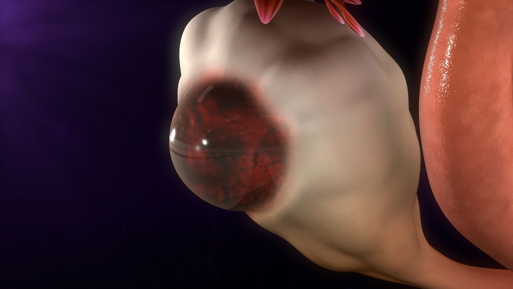
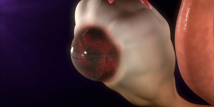
The uterus is the organ that houses the fertilized ovum. It accepts the ovum from the fallopian tube and nurtures it through implantation into the endometrium and fetal development until childbirth. During pregnancy, it also expands to accommodate the developing fetus. The uterus also directs blood flow to the pelvic area and the ovaries through its vascular supply.
The ovary is the other major adnexal structure that makes up a woman’s gynecological system. It can form cysts of different types, which may be benign or malignant. Its blood supply comes from the ovarian artery, which is a direct branch of the aorta. This relationship is important when performing a surgical procedure such as a hysterectomy, where careful clamping of the ovarian artery is required to prevent damage to the ureters (the two branches that drain the urine from the ovaries).
A woman’s uterus has four major regions: the fundus, which is the broad curved upper region; the body, which begins immediately below the fallopian tubes and extends downward; the isthmus, which is a narrow neck region; and the cervix, which stretches downward from the isthmus until it opens into the vagina. The uterus is 6 to 8 cm wide, but it is only about half as wide at the isthmus.
Abnormalities of the uterus can affect any of its three layers, but most common problems target the myometrium and endometrium. Infections of the uterus can cause pain and bleeding during menstrual periods. Abnormalities in the lining of the uterus can cause abnormal vaginal discharge and difficulty with conception. Abnormalities in the uterus can also lead to miscarriage or premature birth.

Uterine abnormalities include a wide range of conditions, from polyps and cysts to cancer and adenocarcinoma. These conditions may be treated with medications, a change in diet or exercise and sometimes surgery.
Other conditions that affect the uterus can be caused by hormones or by pregnancy or childbirth. These include:
Fibroids: Non-cancerous tumors of the muscular tissue of the uterus, also known as leiomyomas or myofibromas. They may be a normal part of the aging process, but can also occur due to genetic factors and hormones or due to certain diseases such as endometriosis or tuberculosis.
Endometrial hyperplasia: A condition where there is an abnormal growth in the lining of the uterus. It can be either stromal or glandular, and can have normal or atypical cells or simple or complex glandular structures. Some kinds of endometrial hyperplasia are at higher risk of becoming cancer.
A woman’s gynecologist can diagnose uterine disorders by taking a detailed medical history, performing a pelvic exam and ordering laboratory tests such as a PAP smear or an ultrasound. Other tests may be needed to determine the cause of your uterine symptoms, such as a CT scan or MRI. Depending on the results, treatment options may include medication (such as hormonal pills or shots) or surgery such as a hysterectomy or myomectomy. In some cases, ‘watchful waiting’ may be recommended, which means you will need regular check-ups and blood tests to monitor the condition and track any changes.


