


Accurate cancer staging is vital for multiple stakeholders, including patients, physicians, and researchers. It can help researchers select patients and stratify them for clinical studies. It can also help epidemiologists track the impact of population-based screening. The following sections provide a brief description of the differences between cancer staging systems. They should be familiar with the limitations and benefits of each. We also recommend using an appropriate staging system based on the characteristics of the patient’s tumor.
Clinical staging determines the stage of cancer based on the size of the tumours within the primary site of origin and the extent of the disease elsewhere in the body. The information gained from cancer staging helps doctors provide the correct prognosis for patients. Accurate staging also helps physicians devise individualized treatment plans for each patient. If the cancer is detected early, treatment options can be tailored to the patient’s condition and stage. However, patients should be aware of the importance of obtaining an accurate diagnosis from the start.
The GCCS meeting, held in London, UK, involved experts from many organizations and reaffirmed the need to develop a consensus on cancer staging terminology. The meeting also included a literature review to understand the reasons behind differences in staging terminology. The participants reaffirmed the importance of cancer staging terminology and agreed on the meaning of the terms used in the process. They also defined relevant terms to improve cancer diagnosis and treatment. These efforts have provided the foundation for an improved understanding of cancer staging.
Although there is no single definitive method for lung cancer staging, the use of multidisciplinary teams provides a collaborative platform for experts in the field. The assessment of mediastinal lymph nodes is an important component of cancer staging. There are several techniques that can help achieve optimal mediastinal staging. However, there are few studies to prove the benefits of multidisciplinary teams. The aim of these teams is to provide personalized treatment and care to patients with lung cancer.
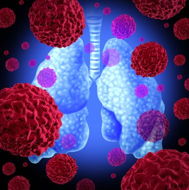
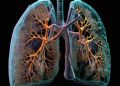
The two most common techniques for determining lung cancer staging are PET-CT scans and CT. PET-CT is the preferred imaging technique for lung cancer and is compared to CT. However, it reduces the radiation dose by 30 percent. Further, it allows doctors to determine the best treatments for a patient. These techniques also have the added benefit of being more accurate and less invasive than conventional methods. For these reasons, cancer staging in lung disease has become an essential part of clinical care.
In some cases, anatomical locations may be difficult to visualize and detect. Because of these differences, tumors may be misstaged. For instance, a T1 lesion may be properly classified as a T2 lesion if the invasiveness of the cancer is limited to the vermilion lip. However, tumors that spread from the mouth or ear may not be diagnosed until they spread throughout the body. If a patient experiences a recurrence of the disease, treatment can be modified to minimize the risk of the cancer.


