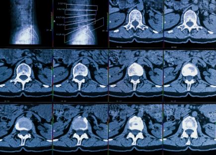
The vertebral column is made up of 33 bones grouped into divisions called cervical (neck), thoracic (upper back), and lumbar (lower back). Each of these bone segments is connected to the next by an intervertebral disc, a tough fibrous cushion with a soft cartilage core.
In a healthy spine, the discs allow for normal motion without compressing nerves or causing pain. However, as people get older, the discs can dry out or become brittle and thin. They may also crack or herniate through the tough outer layer, leaving a hole that can pinch a nerve and cause pain.
Degenerative diseases of the vertebra symptoms vary from person to person but typically include pain, weakness, numbness, and loss of strength. Symptoms can be severe and interfere with daily activities.
Depending on the type and location of the deteriorating disc, your doctor might prescribe anti-inflammatory or painkilling medication to help relieve the pain. Your doctor may also recommend physical therapy and special exercises to strengthen muscles in the back and neck and increase flexibility.
Muscle relaxant medications can also be used to ease pain and muscle spasms caused by slipping and herniated discs. These medications may be given for a short time, then you may stop taking them to see if the pain goes away on its own.
A variety of diagnostic tests can be used to find out the exact cause of your spinal problems and the best course of treatment. These tests include X-rays, magnetic resonance imaging (MRI), and computed tomography (CT) scans.

Your healthcare provider will also do a physical exam to look at the shape and size of your spinal bone and check for any injury or fractures. You might be asked to lie on a table, bend forward, and twist to see how much pain you feel. You may also be asked to touch or press on specific areas of your back and leg to determine if your muscles respond appropriately to pressure.
If the doctor is unable to identify the cause of your pain, you might be tested for spinal stenosis, a narrowing of the spinal canal and nerve roots caused by a herniated or bulging disc. This test involves injecting a dye into the spinal fluid and taking several X-rays.
When spinal stenosis is found, your doctor might recommend a surgical procedure called a spinal fusion. This fusion is performed by inserting a small piece of bone graft between two vertebrae to create a permanent space between the vertebrae, alleviating pressure on the spinal cord and nerves.
Spinal fusions are usually done for the most part conservatively, but some patients may require surgery to remove herniated or bulging discs that are causing pain. During the surgery, the doctor will replace the damaged disc with an artificial one that will provide more stability to your spine.
The spine is a complex system of joints, ligaments and muscles that spreads load across many sections. When disease interrupts this system, focal loads increase in adjacent segments that must move more dramatically to compensate for less movement at the diseased segment.


