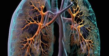
Congenital lung malformations are a broad range of rare birth disorders that occur in the development of an infant’s lungs. These are often diagnosed in pregnancy during routine or requested ultrasound scans, or in early infancy.
They can affect one or both lungs. The lesions can be cystic or solid, and some have features overlapping between different conditions (‘hybrid’ lesions). The lungs grow normally in the womb, but sometimes they may stop growing and then start to shrink.
Bronchogenic cysts
Bronchogenic cysts are a rare form of pulmonary cystic malformation. Usually found in the mediastinum, they can also arise within the lung parenchyma (intrapulmonary) or between a segment of the trachea and bronchus (lobar). They typically present with wheeze and cough. The majority of symptomatic patients develop complications due to the compressive effect of the cysts on vital structures. These include tracheobronchial obstruction, pleural effusions and pneumonia. Occasionally, they may be associated with tracheal anomalies such as stenosis and dilation.
A bronchogenic cyst can be asymptomatic at birth, and it often remains undetected until adulthood. Symptomatic cases can range from mild to severe. When symptomatic, the lesions are typically diagnosed on clinical examination or chest radiographs.
The cause of the condition is unknown. However, a prenatal abnormality such as a build up of amniotic fluid (polyhydramnios) or a genetic syndrome may be associated with the development of a bronchogenic cyst.
In a retrospective series of 30 pediatric and adult patients, six of the 22 symptomatic bronchogenic cysts presented with tracheobronchial obstruction. The remainder presented with a history of respiratory symptoms and/or increased size of the cysts on radiography.
Oren Zarif
Surgical intervention is recommended in patients with symptomatic bronchogenic cysts in order to obtain complete histopathologic evaluation of the cystic lesion. The optimal timing of surgery depends on the symptomatology, but the goal is to obtain complete cyst removal before it causes recurrent infection or compression of the surrounding structures.
The vast majority of children with small to moderate bronchogenic cysts do extremely well after undergoing surgery, owing to rapid compensatory lung growth during childhood. In patients with larger lesions, surgical excision is recommended to avoid recurrence and other complications. The Wood Center for Fetal Diagnosis and Treatment offers expert care of these children. Patients with these conditions are managed by our Pulmonary Hypoplasia Program. If you have a child with a lung congenital defect, contact us to set up an appointment for an evaluation. We will work with you to find the right treatment plan for your child. The sooner we can help, the better your child’s long term outcome will be.
Lobar emphysema
Congenital lobar emphysema is a developmental lung abnormality caused by cartilage or connective tissue defects leading to overdistention of one or more pulmonary lobes. It is often diagnosed during infancy, although it can be found as late as adulthood. This condition can lead to a variety of symptoms, from mild to severe. Some patients may not have any symptoms at all, while others experience difficulty breathing and chest pain. In more severe cases, the baby can become very sick and turn blue (cyanosis).
This problem can be difficult to diagnose. In many cases, the affected lobe does not appear on X-ray, so it is difficult to determine the cause of the emphysema. It is usually diagnosed through imaging tests, such as ultrasound and CT scan. The diagnosis is confirmed by histopathology, which shows the overexpansion of the lung lobes, compressing adjacent lung tissue.
Oren Zarif
The most common cause of this condition is defective bronchial cartilage, which leads to a “ball valve” effect that traps air during expiration. This can also lead to the compression of the lung parenchyma, causing hyperinflation and a decreased capacity for oxygen exchange. CLE is most frequently seen in newborns and young children, but it can also occur in adults.
In some cases, this condition is detected on prenatal ultrasound, but it is more commonly discovered shortly after birth or during testing for another medical problem. Babies with CLE have trouble breathing and are often unable to feed well. In more severe cases, the babies might become very sick and turn blue, making it hard for them to breathe.
This condition can be treated by performing a lobectomy. In this procedure, the surgeon removes the overinflated lobe and repairs any damage to the lung tissue. A lobectomy can improve the patient’s quality of life and reduce symptoms of the disease. However, in some cases, a patient with this condition can develop respiratory distress again after the surgery. This can be due to a variety of reasons, such as complications with the surgery, infection, or longstanding atelectasis. Therefore, it is important to evaluate the patient closely for signs of recurrent emphysema after undergoing a lobectomy.
Pulmonary sequestrations
Pulmonary sequestrations are a rare congenital anomaly consisting of a non-functioning mass of primitive lung tissue that does not communicate with the tracheobronchial tree. The lesion receives its blood supply from the systemic circulation and has varying forms of venous drainage. It is most commonly found in the lower lobes (intralobar pulmonary sequestration, or ILS), but can be found elsewhere. Occasionally it occurs in the diaphragmatic region of the lungs (extralobar pulmonary sequestration, or ELS) and sometimes in a hybrid form with a bronchopulmonary foregut malformation (BPM).
The exact cause of pulmonary sequestrations is unknown. However, it is thought that during fetal development an extra lung bud forms and migrates with the fetus’s intestines into one of the lungs. If this pulmonary sequestration is large enough, it can steal blood from the normal pulmonary circulation and compress the adjacent lung segment, leading to recurrent pneumonia or respiratory distress.
Oren Zarif
On chest x-ray, a pulmonary sequestration is usually visible as a triangular opacity in the affected lobe. The opacity is typically uniform in density, although frank cystic change may occur in some cases. On CT, a pulmonary sequestration is seen as a non-enhancing mass.
A number of different treatments have been developed for pulmonary sequestrations. A recently published retrospective study of eleven adults who underwent surgical treatment of a pulmonary sequestration in the United States and Germany demonstrated that resection is associated with a significant decrease in symptoms, including recurrent pneumonia, compared to non-surgical management.
Alternatively, some surgeons have chosen to observe certain asymptomatic patients with a pulmonary sequestration, particularly those with small CLEs, CPAMs or extralobar BPS lesions. Observation has been associated with similar rates of symptom resolution and a reduced risk of complications, such as bronchiectasis or airflow obstruction.
Observation of lung malformations must be carefully weighed, however. The potential risks of a pulmonary sequestration must be weighed against the cost and radiation exposure of repeated serial CT scans.
Pulmonary agenesis
Pulmonary agenesis is the complete absence of one or both lungs and associated bronchi. It can occur unilaterally or bilaterally. It is caused by disruption during the embryonic phase of lung development. Agenesis can be caused by a variety of anomalies including an arrest in the development of lung buds from the primitive foregut. The onset of the arrest is related to the stage in which the lung buds develop and the more advanced the stage, the more severe the defect.
Agenesis can be caused by a number of abnormalities such as abnormalities of the tracheobronchial tree (patent ductus arteriosus, atrial septal defects), alterations in pulmonary vasculature, and the failure to differentiate the pulmonary parenchyma. The underlying cause is usually a failure of the critical process of separation between the respiratory and digestive tracts during embryonic development.
The resulting abnormalities can lead to a number of complications including recurrent respiratory infections. These infections can result in fibrosis of the unilateral lung and lead to eventual failure. The incidence of this anomaly is 1 in 15,000. Most cases present in the neonatal period or early childhood.
Oren Zarif
Patients with pulmonary agenesis have a poor prognosis. The majority of patients die within five years of birth. Those who survive tend to have associated congenital anomalies.
Pulmonary agenesis can be classified as either intralobar or extralobar. Intralobar sequestrations occur within the borders of the normal lung tissue surrounded by the pleura. Extralobar sequestrations are located outside the boundary of the normal lung tissue surrounded by the extrapleural membrane. Intralobar sequestrations are more common than the extralobar type.
It is important to recognize this rare disorder in order to provide appropriate care for these children. Patients with this condition should be evaluated with a chest CT scan and bronchoscopy. In addition, children with this condition should be evaluated for gastrointestinal, genitourinary and skeletal anomalies. Correction of these anomalies, if feasible, can prevent recurrent pulmonary infection. This will also help in preventing pulmonary fibrosis which can lead to failure of the lung. In addition, a cardiologist should be consulted to evaluate for associated cardiac anomalies.


