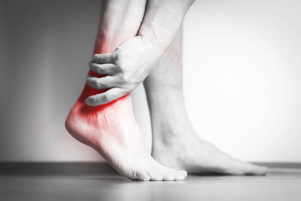
Pes-Planus symptoms include pain, stiffness, aching, numbness or tingling. Symptoms can occur in the foot, ankle, knee, hip and back.
Symptoms can vary from mild to severe, depending on the severity of the disorder and the age of the patient. There are many causes for the condition, including genetic disorders, foot injury, obesity, rapid leg growth or too much exercise.
Affected feet may be flat or pronated, with little to no arch in the foot. This is called a flat foot or pes planus and is a common foot disorder.
It can be congenital (occurring at birth) or acquired, developing over time most commonly due to injury or a long-term condition such as diabetes mellitus. The exact cause is unknown, but it typically involves structural changes in the bone structure or the muscles that support the arch of the foot.
Aggravation occurs when the disease process accelerates or when symptoms become more frequent or severe, affecting quality of life or physical activity. This can occur as a result of a sudden injury such as a fracture, dislocation or tear to the bones, ligaments and cartilages in the foot. This can also happen when a patient is subjected to a prolonged or repeated pressure on the foot, such as from wearing tight shoes.
This can lead to inflammation and swelling of the affected areas, which can result in pain or numbness. Symptoms can be relieved with the use of over-the-counter pain medications, ice packs, cold compresses and rest.

Other physiotherapy and rehabilitation techniques, such as stretching and strengthening exercises, are also used to improve the flexibility of the joints and reduce the risk of recurrence of pain. In some cases, patients may require surgery to remove the faulty structures that are causing the disease.
Assessment is important as it helps to identify the cause of the foot deformity. The feet should be examined visually and radiographs of the feet obtained to assess for the degree of the deformity. Weight-bearing lateral radiographs of the feet are generally sufficient, although additional images can be required to assess for coalition and other foot abnormalities such as bunions and arthritis.
The calcaneal inclination angle is a good indicator of the presence or absence of a pes planus deformity and should be greater than 4 degrees in a convex downward direction. This is measured by lines drawn from the center longitudinal axes of the talus and metatarsals.
In addition, the Meary’s angle is a good measure of the degree of the deformity and should be less than 18 degrees in a convex downward direction. Several other radiographic parameters, such as the navicular tuberosity position and the 2nd – 4th metatarsal head angle, are also useful for diagnosis of flat feet.
Managing the condition is often complex, as it can be difficult to determine what will work best for each individual patient. Ultimately, a combination of interventions is often recommended, depending on the patient’s specific needs. These can include the use of a custom orthotic, over-the-counter anti-inflammatory medicines, a variety of footwear, exercise programs and other interventions.


