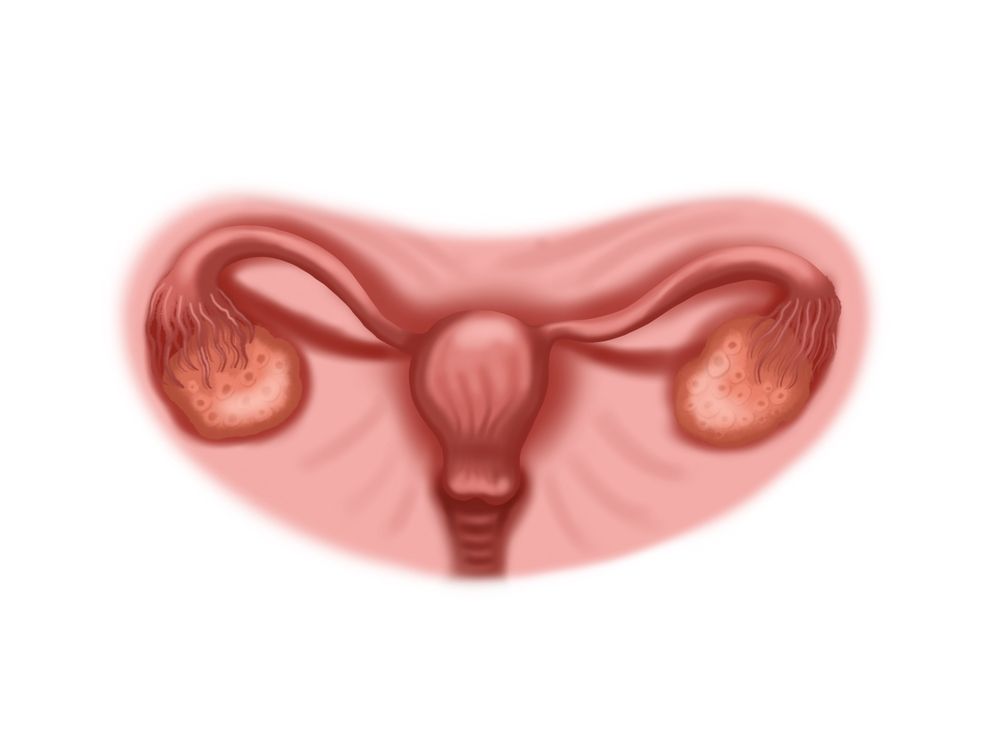
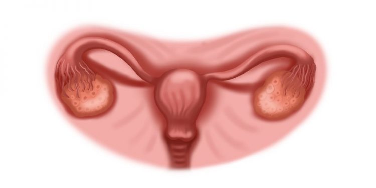
The uterine tube (also known as the fallopian tube, Mullerian duct, and the vaginal canal) is the female structure that transports the ova from the ovary to the uterus each month for fertilization and implantation. The uterine tube is also part of the reproductive system, and the ovaries are attached to it by ligaments at each end.
The human uterine tube develops from paramesonephric (also called Müllerian) ducts in the early embryo. The ducts are formed from the mesoderm, one of three germ layers that form the embryo, which later undergo elongation and coiling to become fully developed uterine tubes. The uterine tube is surrounded by peritoneum, which forms a mesentery for the ovary at each end and contains the blood vessels that enter and supply it.
An opening, the tubal ostium, projects into the abdominal cavity at the site of the pelvic brim and the external iliac artery, which pass downward and cross the infundibulopelvic fold of peritoneum to reach the ilium. From there they curve upward, traverse the adnexa of the uterus, and reach each ovary at the tubal pole, where they are supported by a suspensory ligament (the infundibulopelvic ligament or ovarian ligament).
A thin-walled portion of the tubal lumen, the isthmus, forms more than half of the length of the fallopian tube and is about 1 cm wide; it connects the infundibulum to the more proximal part of the tubal body, which is the intramural or interstitial portion of the uterine tube. The tubal body then reaches the uterus through a narrow, slitlike portion of the uterine wall, and opens into the uterine cavity near the free border and medial surface of the ovary. At the ends of the tubal lumen are a pair of fimbriae. The fimbriae attach to the ovary and extend over it; when an ovum is released in the tubal lumen, it moves through the fimbriae and over the ovary by peristaltic contraction of smooth muscles of the tubal walls, assisted by tubal cilia and secretions.

If the oviduct is obstructed by an abnormality of the ovary or tubal structures, sperm may reach and fertilize the ovum in the anterum portion of the tube and implant it there instead of in the uterus, resulting in an ectopic pregnancy. The ectopic pregnancy can only be cured by surgical interference, and it is potentially life-threatening because of its proximity to the internal os.
An occluded uterine tube can be reopened by tubal ligation, a procedure that involves surgically cutting and severing the intramural or interstitial portion. However, the ovary is attached to the occipital end of the tubal tube by a ligament; this attachment can lead to complications such as salpingitis, when fibrous adhesions, or hydatid cysts, are present. An alternative to tubal ligation is the use of a P-block device, which consists of a nylon skeleton with a hydrogel body that expands and occludes the uterotubal junction after insertion into the uterus.


